Liver fluke infections in ruminant livestock are still significantly prevalent. During inspections of sacrificial animals, liver fluke infections are common and routine. Even in 2026, numerous reports of liver fluke infections in sacrificial animals continued. Post-mortem examinations, or examinations after animal slaughter, still reveal cases of liver fluke. Although the number of cases remains quite high, the severity varies, ranging from mild, moderate, to quite severe.
This situation indicates that livestock farmers vary in their care and treatment practices. The low infection rate indicates that farmers are beginning to pay attention to the health of their livestock. Some farmers have been administering regular anthelmintic treatments. However, because these cases are still common, we need to improve our husbandry practices, particularly feeding schedules and disease prevention. One high-risk habit is mowing grass too early in the morning, when the dew is still present, as worm eggs or larvae often cling to the tips of the grass at that time.

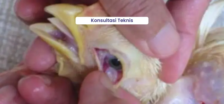
Liver Fluke Infection Findings
Early diagnosis of liver fluke disease requires examination, clinical symptoms, liver examination, and laboratory tests. In addition to laboratory tests, farmers should be alert to any physical changes in their livestock, especially if the infection has progressed. Symptoms may include:
- Stool excretion disorder: Livestock have difficulty defecating with dry droppings, or in chronic cases, have continuous diarrhea.
- Stunted growth: Livestock appear smaller than the herd or their growth is slow even though feed is sufficient
- Decrease in productivity: Daily weight targets were not achieved and livestock productivity declined drastically.
Often, in the mild stage, livestock appear healthy, but their livers are beginning to deteriorate. This is why regular examinations are essential. Post-mortem examinations can only be performed on animals that have already died or been slaughtered. These examinations will reveal adult worm infestation in the liver, liver damage, and thickening of the bile ducts.

Laboratory testing through stool examination is a crucial step in establishing an accurate diagnosis by identifying the presence of worm eggs microscopically. The advantage of this method lies in its ability to detect infections from the early (mild) to chronic stages, allowing for more effective and targeted treatment.
Any liver where liver flukes are found is unfit for consumption. Infected organs must be removed immediately to prevent health risks. Health teams will typically conduct pre- and post-slaughter inspections to ensure the safety of the public and consumers.
The causes of liver fluke infection in cattle are Fasciola gigantica and Fasciola hepatica. Cattle become infected when they eat grass contaminated with worm larvae (metacercariae) that are found in their feed or drinking water. Worm eggs are shed in the feces of infected cattle. In a moist environment, the eggs hatch and infect freshwater snails (which act as temporary hosts). After developing, the larvae emerge from the snails and attach to grass. Cattle that eat grass contaminated with these infective larvae will migrate through the intestinal wall to the liver and bile ducts to develop into adult worms. These adult worms can then damage the liver.
Prevention of Liver Fluke Infection
Before liver fluke infection occurs, farmers can implement a preventative program to prevent losses. Some of the potential negative impacts include weight loss, stunted growth, decreased meat and offal quality, decreased milk production, and the risk of human transmission (zoonosis). To avoid these, the following measures can be taken:
- Giving worm medicine to eradicate liver flukes with Wormzol Suspension upon arrival of the stock. If maintenance is longer, it may need to be repeated every 3-4 months.
- Prevent the cage from becoming wet, feces piling up, and damp. Routinely sanitize the cage and equipment by cleaning, washing, and spraying with disinfectant (Neo Antisep, Medisep) every day.
- Eradication of the temporary host, namely freshwater snails.
- Avoid cutting grass at least 30 cm from the ground surface and do so when there is sunlight or no dew. The grass should be dried by drying it in the sun.
- Avoid grazing livestock in the morning, so that livestock do not consume wet grass tips which may contain metacercariae.
- Monitoring worm eggs and larvae by conducting routine fecal examinations every 2-3 months so that when worm eggs are found in the feces, they can be detected early.